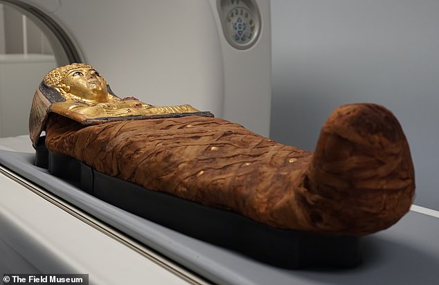
X-ray scans have finally answered the mystery of how a famous Egyptian mummy was placed inside a coffin with no opening.
Scientists were able to see into Lady Chenet-aa’s sarcophagus for the first time to finally understand how she was embalmed 3,000 years ago.
For years, researchers looked for a cavity opening, finding only a tiny gap near her feet that wouldn’t have been big enough to fit her body.
A lack of visible openings or entry points in Chenet-aa’s coffin had previously left researchers unable to access her remains, but new CT scans have removed some of the questions that have plagued scientists for decades.
When the team ran the coffin through the CT scanner, they were able to more thoroughly view the underside, where ‘you can start to see that there’s a seam going down the back and some lacing,’ said JP Brown, Senior Conservator of Anthropology.
Researchers at the Field Museum in Chicago estimated that she was in her late 30s or early 40s when she died and the detailed efforts to prepare her for the afterlife confirmed she was of high social standing.
The new scans revealed that Chenet-aa lived in the Third Intermediate Period of Egypt, during the 22nd Dynasty and is still in ‘remarkable condition.’
The team was also able to discern information about her age, diet and how embalmers prepared her for the afterlife.

New CT scans of Lady Chenet-aa have allowed researchers to gain insight into her age, diet and how she was embalmed

The scans created 3D images of Chenet-aa’s body alongside a second mummy called Harwa who was also considered a high-status individual at the time
The researchers studied two mummies out of 26 that were on display at the Field Museum in September to find out how they were prepared for the afterlife.
Using 3D-scanning technology, they created X-rays of the skeletons and artifacts that were contained inside the coffins for thousands of years.
The most unusual aspect of Lady Chenet-aa’s coffin was the fact that there didn’t appear to have any seams or openings that would have allowed embalmers to place her body inside.
However, the 3D scans revealed that rather than the cartonnage being constructed around the body, as was usual for ancient Egyptians, the coffin was created first and then softened using humidity so it was flexible and could be molded around Chenet-aa.
The embalmers placed stuffing in her trachea so it wouldn’t collapse when she was stood upright, while a slit was cut from the top to the bottom of the coffin and placed over her wrapped body.
After they laced down the back of the coffin, they placed a wood panel at her feet and pegged it in place to keep her from moving while they molded the coffin.
‘From an archeological perspective, it is incredibly rare that you get to investigate or view history from the perspective of a single individual,’ Drake said.
‘This is a really great way for us to look at who these people were – not just the stuff that they made and the stories that we have concocted about them, but the actual individuals that were living at this time.’

The 3D renderings showed that artificial eyes were placed in Chenet-aa’s eye sockets and the sons of Horus were placed in her body cavity alongside her organs

The CT scans took three days to complete but researchers said it could take up to three years before they can fully review the images
They also noticed that Chenet-aa was wrapped in layers of expensive linen and placed in a decorated cartonnage coffin – a type of brightly painted molding similar to papier mache that was reserved for elite burials.
Unlike other mummies whose internal organs were placed in separate jars with the four sons of Horus on the lids, embalmers took a different approach for Chenet-aa.
Scans showed that in her case, the embalmers created packets that the organs were placed into alongside six wax statues of the sons of Horus that were reinserted into the body cavity.
The four sons of Horus included Imsety, who was the human-headed god who protected the liver and Hapy who had the head of a baboon and protected the lungs.
The second two were identified as Duamutef, who had a jackal head and was designated to protect the stomach while the falcon-headed Qebehsenuef protected the intestines.
Images showed she had lost several teeth and the remaining ones had signs of significant wear, indicating that her food contained grains of sand that wore down the enamel.
It also revealed that the embalmers replaced her eyes with artificial ones to ensure they didn’t deteriorate over time.
‘The Ancient Egyptian view of the afterlife is similar to our ideas about retirement savings. It’s something you prepare for, put money aside for all the way through your life, and hope you’ve got enough at the end to really enjoy yourself,’ said Brown.
‘The additions are very literal. If you want eyes, then there needs to be physical eyes, or at least some physical allusion to eyes.’

The scans revealed that embalmers had laced Chenet-aa inside the coffin rather than molding it around her body
The researchers also focused on a second mummified individual known as Harwa who lived around the same time as Chenet-aa and is also believed to be of high social status.
3D images of his spine showed that he didn’t suffer from any ailments related to physical labor and his well-kept teeth reflected that he had access to quality food that was only available to the elites.
The primary goal of their research was to show that the mummies were people and aren’t just objects of art, according to the museum.
‘We are trying to understand them as people so that we can share those stories and insights with the general public to kind of rehumanize and shift the narratives to be more respectful and give some more dignity to these mummified individuals,’ Drake told CNN.
The scans took about four days, however, researchers said a full analysis of their findings could take up to three years to complete.
This article was originally published by a www.dailymail.co.uk . Read the Original article here. .


